Real-Time Proliferation Assays
Cell proliferation is fundamental to many biological processes. It drives organismal growth and tissue repair, while its dysregulation is linked to diseases such as autoimmunity and cancer. Proliferation is also widely used as a general indicator of cell health—for example, to detect viral infection or assess drug toxicity.
Traditional proliferation assays, however, require extensive manual handling and provide only limited endpoint data.
The xCELLigence Real-Time Cell Analysis (RTCA) platform offers a more comprehensive way to monitor proliferation continuously and label-free.
Key Benefits
Direct, real-time measurement of cell health and proliferation
Quantitative, reproducible data
Simple, high-throughput workflow
Deeper insight into cellular mechanisms of action
Multiple Perspectives on Cell Health For Higher Confidence
Conventional assays offer only snapshots. In contrast, the xCELLigence RTCA eSight combines electrical impedance with live-cell imaging to deliver complementary, real-time information on cell number, adhesion, and morphology—without adding workload. This dual-mode approach provides primary and confirmatory results within the same assay.
Application Note: Real-Time Monitoring of Cell Proliferation and Viability
Application Note: Real-Time Monitoring of Apoptotic Cell Death
Multiplexing Impedance with Live-Cell Imaging
xCELLigence impedance technology detects the earliest stages of proliferation by sensitively measuring changes in cell attachment. While imaging-based percent confluence and impedance trends align over long experiments, impedance can detect and rank the effects of different serum concentrations within the first 10 hours—crucial for applications such as cell therapy manufacturing, where early quality attributes matter.
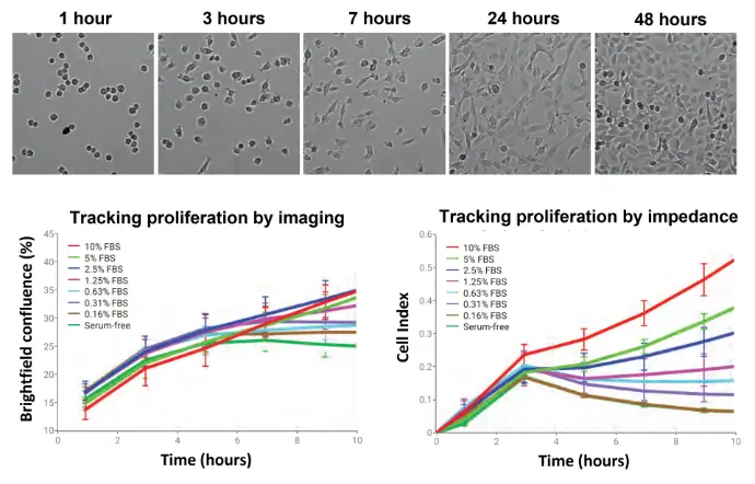
Label-free monitoring of HT-1080 cell proliferation using xCELLigence RTCA eSight. Top images illustrate cell adhesion, spreading, and growth over time, while bottom graphs show proliferation kinetics at different serum concentrations using % brightfield confluence or impedance. Notably, impedance detects and rank-orders serum-dependent effects within just 10 hours, providing a highly sensitive early readout.
Tracking HT-1080 proliferation using nuclear-localized fluorescent eLive dyes to obtain actual cell counts. Top images show cells labeled with eLive Red or eLive Green, with eSight masking (right panels) outlining each nucleus. Bottom panels display (left) proliferation curves based on fluorescent cell counts, (middle) brightfield confluence comparisons with and without eLive dyes, and (right) impedance-based proliferation kinetics. Error bars represent standard deviation across 12 replicate wells.
Tracking Proliferation by Actual Cell Counts Using Fluorescent Dye-Labeled Nuclei
Agilent eLive Red and eLive Green dyes enable direct cell counting on the eSight. When added to culture media, these dyes produce minimal background fluorescence but increase >1,000-fold upon binding to nuclear DNA—allowing accurate, nonperturbing nuclear counts even over multi-day experiments.
Application Note:
Monitoring Cell Proliferation in Real Time
Tracking Proliferation by Actual Cell Counts Using Fluorescent Protein-Labeled Nuclei
Stable fluorescent cell lines can be generated using eLenti Red, eLenti Green, and eLenti Blue lentiviral reagents. These integrate into the host genome and drive expression of nuclear-localized mKate, EGFP, or TagBFP via an EF1α promoter. eSight imaging displays clear nuclei with or without automated fluorescence masking.
Tracking cell proliferation using nuclear-localized fluorescent protein to determine actual cell numbers. Example Images for HEK-293, MDCH, and MDA-MB.231 cells transduced with Agilent eLenti Red, eLenti Green, and eLenti Blue, respectively.
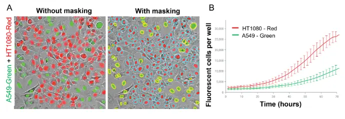
Image-based monitoring of proliferation for distinct cell types within a mixed population of cells. (A) Left: Image of EGFP-expressing A594 cells mixed with mKate-expressing HT1080 cells. Right: Same as left panel but with eSight fluorescent nuclei masking applied (yellow outline around green nuclei and aqua outline around ned nuclei). (B) Proliferation of A549 and HT1080 cells within this coculture context. Error bars reflect standard deviation for 96 replicate wells.
Analyzing Proliferation in Mixed Cell Populations
In a mixed-culture example, EGFP-labeled A549 cells and mKate-labeled HT-1080 cells were seeded at a 1:1 ratio. Using separate red and green nuclear masks, eSight accurately revealed the proliferative advantage of HT-1080 cells. This same method can be applied to studies involving the tumor microenvironment, co-cultures, or immune cell–mediated killing.
Literature
Monitoring Cell Proliferation in Real Time Using the xCELLigence RTCA eSight
Real-Time and Dynamic Monitoring of Cell Proliferation and Viability for Adherent Cells
Combining Live Cell Imaging with Cellular Impedance to Monitor Apoptotic Cell Death in Real Time
For Research Use Only. Not for use in diagnostic procedures.