3D Cell Culture: Form, Grow and Analyze Spheroids and Organoids
Form spheroids, keep organoids in long-term culture, analyze and isolate them: 3D cell culture tools and advice from one source.
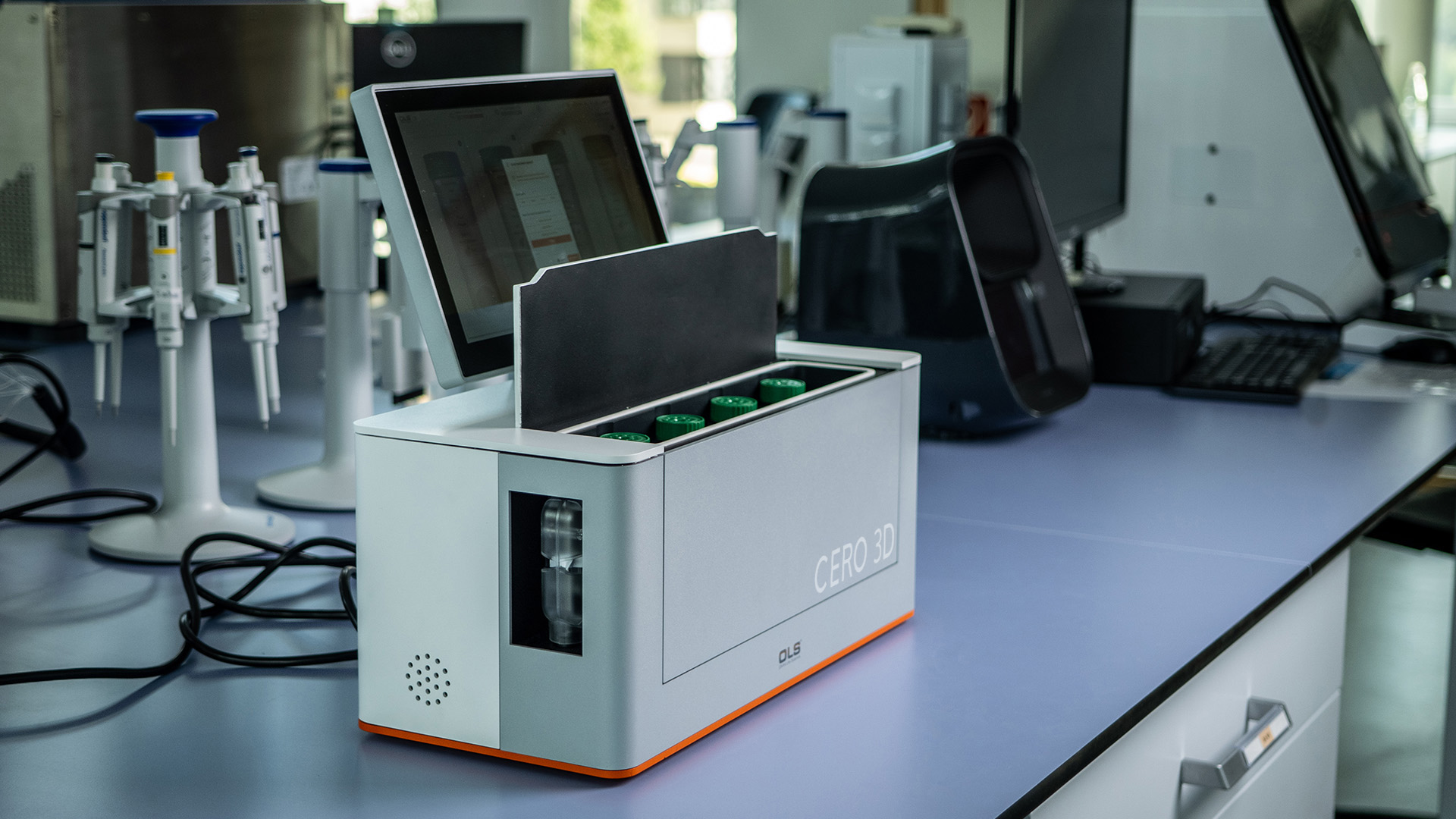
Why 3D Cell Culture? Where 2D Models Fall Short
Cells grown as a flat monolayer on plastic lack what defines real tissue: a three-dimensional architecture, contacts to neighboring cells and to the extracellular matrix, and gradients of nutrients and oxygen. That is why 2D models often predict drug efficacy and toxicity poorly.
In 3D cell culture, cells grow as spheroids, organoids or tissue pieces. These models reproduce cell-cell and cell-matrix interactions, oxygen gradients with hypoxic cores, drug penetration and resistance as well as the formation of extracellular matrix. That makes them more predictive for tumor biology, toxicology, virology and regenerative medicine.
3D culture is more demanding, though. Typical challenges are:
- heterogeneous cell populations when pluripotent stem cells are differentiated,
- loss of structure and metabolic activity of organoids in long-term culture,
- time-consuming manual maintenance such as media changes,
- high costs for consumables and substrates.
The tools below address exactly these points – from uniform spheroid formation to long-term culture, real-time analysis and the isolation of single organoids.
Spheroids, Organoids or Tissue: Which 3D Model Do You Need?
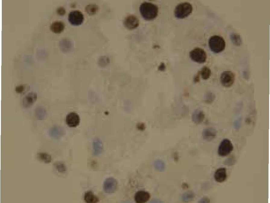
Spheroids
Compact aggregates of cell lines or primary cells – simple to form and well suited to tumor biology, drug and toxicity testing and virology. Examples from research labs: HepG2 spheroids kept for more than 80 days in CERO 3D, myospheres from porcine primary cells and intestinal tumor spheroids for nanoparticle testing.
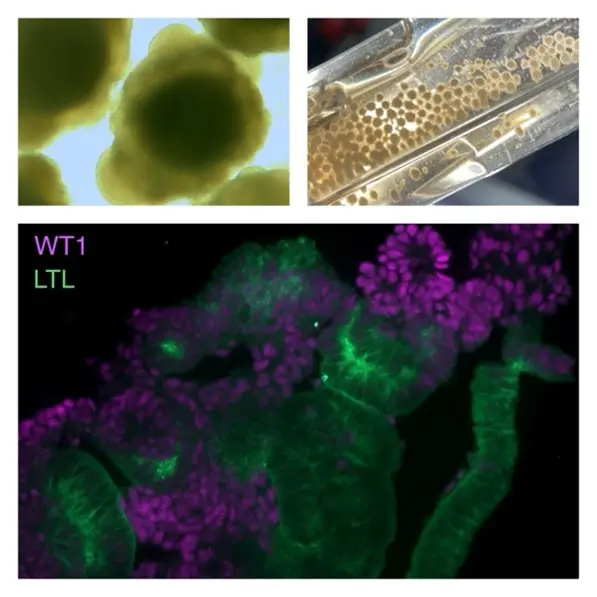
Organoids
Self-organized mini-organs from primary tissue, embryonic stem cells or iPSC that reproduce key features of organ structure and function. Examples: gastric organoids (Technical University of Munich), kidney organoids (Nephrolab Cologne), cerebral organoids and glioma assembloids (Sevenich lab) and hematopoietic organoids for CAR macrophages.
Stem Cells and Tissue
Expand induced pluripotent stem cells as 3D aggregates or on microcarriers, differentiate them into beating cardiac bodies and other tissue models, or keep tissue biopsies in long-term culture.
Scaffold-Free or Scaffold-Based: Methods for 3D Cell Culture
Scaffold-Free
Cells aggregate on their own, without an embedding substrate. Common formats are ultra-low attachment plates, hanging drops and dynamic suspension culture in a bioreactor. Scaffold-free methods yield dense aggregates and are easy to handle.
From OLS: spheroid plates (CEROplate ULA) and the 3D cell culture bioreactor CERO 3D.
Scaffold-Based
Cells grow in or on a matrix – for example in hydrogels such as collagen or basement membrane extracts, or on microcarriers. The scaffold gives you control over stiffness and environment but adds a substrate to the workflow.
From OLS: alginate microcarriers with adjustable stiffness (2–8 kPa) and size (100–500 µm).
Your 3D Workflow: Form, Grow, Expand, Analyze, Isolate
From the first spheroid to the isolated organoid – a matching tool for each step, and advice from one team.
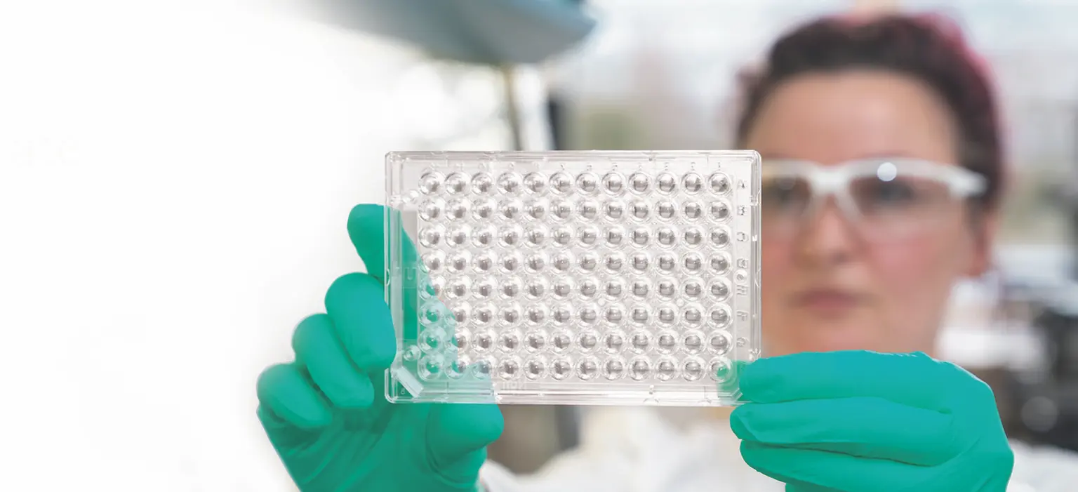
Form: CEROplate ULA Plates
Uniform spheroids within 2–24 hours, depending on the cell type. U-bottom 96- and 384-well plates hold one spheroid per well; 6- and 24-well plates have a flat bottom. Sterile, endotoxin-free and non-cytotoxic – formation and analysis run in the same plate.

Grow: CERO 3D Incubator & Bioreactor
Scaffold-free culture with low-shear, bi-directional rotation in four individually controlled CEROtubes. CO₂, temperature and pH are controlled automatically, for cultures lasting from days to months.
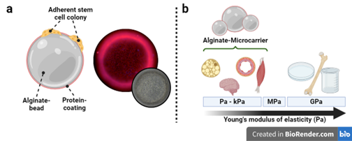
Expand: Alginate Microcarriers
Soft alginate microcarriers for the expansion of hiPSC in the CERO 3D: more than 85 % viability and more than 6 × 10⁶ cells after four days.

Analyze: xCELLigence RTCA eSight
Follow tumor spheroid growth and killing by immune cells in real time – for example with images every 4 hours over 10 days.
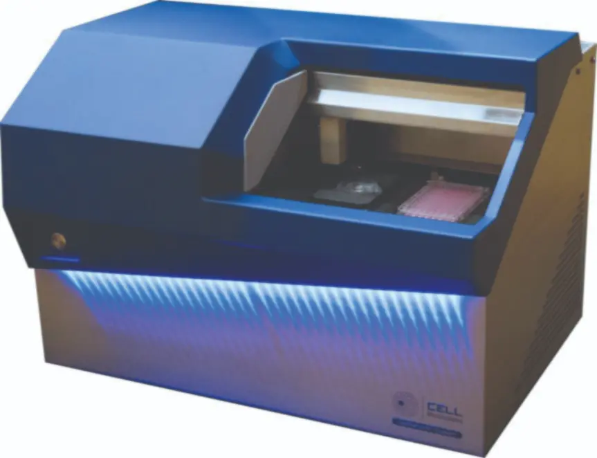
Isolate: CellRaft AIR System
Grow hundreds of organoids on one array, image them as z-stacks in brightfield and fluorescence and retrieve intact organoids automatically from the matrix into 96-well plates for downstream assays.
Feed: cell culture media, sera and supplements from PAN-Biotech, including stem cell media and serum-free media.
ULA Plate or Bioreactor? How to Choose
A ULA plate (CEROplate) fits when you …
- need one spheroid per well,
- test many conditions in parallel, for example for screening in 96- or 384-well format,
- want to form and analyze spheroids in the same plate.
A bioreactor (CERO 3D) fits when you …
- need large numbers – in the Sevenich lab, more than 200 cerebral organoids per CEROtube,
- need cultures to stay stable for a long time – more than 80 days for HepG2 spheroids, more than five months for cerebral organoids,
- want to expand and differentiate stem cells in the same vessel.
In a side-by-side comparison with 96-well ULA plates, glioma assembloids grown in CERO 3D reached a connected surface of more than 50 % instead of less than 10 % and showed less apoptosis. Read the Sevenich lab report.
For kidney organoids, CERO 3D allows direct inoculation, so a separate aggregation step in ULA plates is not needed.
Not sure which approach fits your cells? Ask our specialists.
Proven in Research Labs
More results: published studies with CERO 3D.
Frequently Asked Questions about 3D Cell Culture
In 3D cell culture, cells grow in all three dimensions – as spheroids, organoids or tissue pieces – instead of as a flat monolayer on plastic. This restores cell-cell and cell-matrix contacts, nutrient and oxygen gradients and a tissue-like architecture, so the cells behave more like they do in the body.
2D culture is simple, inexpensive and easy to image, but cells on a flat surface lack the architecture, matrix interactions and gradients of real tissue, which limits how well 2D models predict drug efficacy and toxicity. 3D models close that gap, at the price of more demanding handling, culture and analysis.
Spheroids are compact cell aggregates, usually formed from cell lines or primary cells, and are widely used for tumor, drug and toxicity studies. Organoids are self-organized, miniaturized organ models derived from primary tissue, embryonic stem cells or iPSC; they reproduce key structural and functional features of organs such as brain, gut, kidney or liver.
Scaffold-free methods – ultra-low attachment plates or suspension culture in a bioreactor – let cells aggregate on their own, without embedding substrates, and yield dense aggregates. Scaffold-based methods grow cells in hydrogels or on microcarriers and give you control over stiffness and environment, but add a substrate to the workflow. The right choice depends on your cell type and research question – our specialists are happy to help.
Use ULA plates such as CEROplate when you need one spheroid per well and many conditions in parallel, for example for screening in 96- or 384-well format. Use a bioreactor such as CERO 3D when you need large numbers of organoids or spheroids, long-term culture over weeks to months, or stem cell expansion and differentiation in the same vessel.
In CEROplate ULA plates, uniform spheroids form within 2 to 24 hours, depending on the cell type. In the CERO 3D bioreactor, HepG2 spheroids have been kept viable for more than 80 days and cerebral organoids for more than five months.
CEROplates are compatible with microscopy, fluorescence imaging and automated high-content screening. The xCELLigence RTCA eSight follows spheroid growth and immune-cell killing in real time, and the CellRaft AIR System images hundreds of organoids and retrieves intact ones from the matrix.
Planning a 3D Project?
See CERO 3D in your lab or online – or ask our specialists which approach fits your cells.