Cytotoxicity Assays
Traditional assays for evaluating cytotoxicity typically probe membrane integrity by looking at the passage of labels into the cell or the leakage of biomolecules out of the cell. However, these assays are time consuming and provide only endpoint data. In contrast, the xCELLigence RTCA cytotoxicity assay provides quantitative, real-time data for more detailed insight into cellular mechanisms causing cytotoxicity. Applications for measuring cytotoxicity span from investigating tumor cytotoxicity in 3D cell cultures, antibody dependent cellular cytotoxicity (ADCC), environmental water sample monitoring, to high-throughput drug screening and profiling.
Overcoming Limitations of Traditional Cytotoxicity Assays
Track drug or compound effects on cell health, proliferation, and morphology in real time.
Generate dose-response curves and monitor long-term cellular responses with minimal effort.
Track 2D or 3D tumor cell growth and shrinkage via live cell imaging.
A simple and high-throughput method.
Cytotoxicity Screening and Profiling
Easily perform high-throughput compound screening and profiling with the xCELLigence RTCA HT instrument, which provides real-time monitoring of changes in cell attachment, morphology, and growth characteristics as a response to cytotoxic agents. Make informed decisions about the timing of cell treatments and easily generate dose-response curves at multiple time points, without increasing the workload.

Real-time cytotoxicity profiling of A549 lung carcinoma cells using the xCELLigence RTCA HT system. Normalized Cell Index traces and 72-hour dose-response curves (xCELLigence vs. WST-1) illustrate compound effects and corresponding EC50 values.
Dynamic monitoring of cytotoxic compounds
Using impedance-based, real-time analysis, A549 cells were exposed to cytotoxic agents with distinct mechanisms of action -including 5-fluorouracil, vinblastine, and staurosporine. Each compound produced a unique kinetic signature that reflected its mode of action, drug concentration, exposure duration, and cellular response.
Because these signatures are highly characteristic, they can be used to classify compounds with unknown targets by comparing their kinetic profiles to those of known drugs.
Application Note: Dynamic Monitoring of Cell Proliferation and Viability
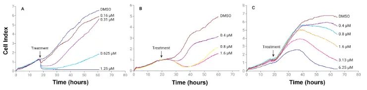
Dynamic monitoring of cytotoxic compounds. A549 cells were seeded in 96X E-Plates and continuously monitored using impedance technology. The cells were treated with the indicated final concentrations of (A) staurosporine, (B) vinblastine, and (C) 5-fluorouracil.
Monitor Cytotoxicity of 3D Tumor Spheroids with xCELLigence RTCA eSight
Monitor and quantify growth (either label or label-free) of 3D single tumor spheroids with Agilent xCELLigence RTCA eSight—all inside your incubator. Perform simple and effective detection of 3D tumor cell growth and shrinkage via live cell imaging utilizing mix and read reagents to fully gauge spheroid proliferation and cytotoxicity. The simple 3D cellular viability or toxicity readouts generate reproducible data, resulting in deeper and more meaningful biological insights into your drug discovery assays.
Investigate 3D Spheroid Cytotoxicity
Tumor spheroid growth is traditionally examined visually by standard microscopy; however, this can be time consuming and labor intensive. The xCELLigence RTCA eSight system provides an image-based high-throughput screening method for 3D tumor spheroids, offering a more automated approach with less hands-on time and simple quantification of tumor growth or shrinkage. With this easy workflow, simply add cell treatments and/or reagents (eLenti, eTox, eAnnexin, or eCapase) at day 3 for quantitative analysis.


Visualize and Quantify Kinetics of 3D Tumor Spheroid Cytotoxicity
In this example, the RTCA eSight live cell imaging system monitors camptothecin-mediated HT-1080 Red spheroids shrinkage. Images show HT-1080 Red spheroids treated by different concentrations of camptothecin and vehicle control for 0, 2, 4, 6, 8, and 10 days. Quantitative analysis of concentration-dependent cytotoxicity effect of camptothecin on HT-1080 Red spheroids is shown. Green fluorescent dead cell marker (eTox Green) and its corresponding segmentation mask (yellow) are also shown.
Application Note: Monitoring Drug-Mediated 3D Tumor Spheroid Shrinkage
Assessing Water Cytotoxicity Using xCELLigence RTCA
See how Alberta Centre for Toxicology scientists developed a label-free, real-time cellular assay for source water monitoring. The scientists collected a total of 436 river water samples for assessment of water cytotoxicity. The assay used HepG2 (human hepatocarcinoma) cells and cytotoxicity response was tested at dilutions of 80%, 60%, 40%, 30%, 20%, and 10% of the original water samples. The Agilent xCELLigence RTCA system monitored HepG2 cell growth following exposure to the combined toxic effect present in water samples.

Literature
Application Notes
Real-Time and Dynamic Monitoring of Cell Proliferation and Viability for Adherent Cells
Monitoring Drug-Mediated 3D Tumor Spheroid Shrinkage in Real Time
Assessing Water Cytotoxicity with an Impedance-Based, Real-Time, and Label-Free Cellular Assay
For Research Use Only. Not for use in diagnostic procedures.