HPV E6/E7 mRNA Expression Analysis Using the Agilent NovoCyte Flow Cytometer
Abstract
The presence of human papillomavirus (HPV) mRNA in a cell indicates viral activity. This application note demonstrates the detection of mRNA for two HPV oncoproteins, E6 and E7, in HPV control cell lines and in cervical cells samples using the FLOWSCRIPT HPV E6/E7 assay and the Agilent NovoCyte flow cytometer.
Authors
Lauren Jachimowicz and Garret Guenther Agilent Technologies, Inc. San Diego, CA, USA
Introduction
HPV is the most common sexually transmitted disease and the chief cause of cervical cancer. HPV infects cervical tissues as well as other mucosal and cutaneous regions of the body. Primarily, HPV infection is asymptomatic and transient, but it may persist and cause cervical cellular abnormalities.
Since the discovery of HPV as the
underlying cause of cervical cancer,
morphologically abnormal cervical cells
have routinely been screened for the
presence of HPV DNA by PCR analysis.
Fluorescent in situ hybridization (FISH) is a powerful technique used for the detection of RNA or DNA in intact cells with a fluorescent probe. Recently, FISH technology has been combined with flow cytometry to allow for rapid examination of RNA expression in individual cells. Detection of viral HPV mRNA instead of DNA is a more direct indication of viral activity; therefore, the detection of HPV RNA is useful for HPV detection. Overexpression of two HPV oncoproteins, E6 and E7, contributes to the malignancy of HPV-infected cells by inhibition and degradation of tumor suppressor proteins, and promotes cell growth.
With innovations in FISH and flow
cytometry, the ability to detect the
presence of E6/E7 HPV mRNA using the
FLOWSCRIPT HPV E6/E7 assay by Enzo
Life Sciences and the NovoCyte flow
cytometer, provides relevant information
at the single-cell level. To demonstrate
the ability of HPV mRNA detection on
the NovoCyte flow cytometer, both HPV
control cell lines and cervical cells were
assayed for E6/E7 mRNA transcription
and analyzed on the NovoCyte flow
cytometer.
HPV RNA detection in cultured cells with the NovoCyte flow cytometer
The FLOWSCRIPT HPV E6/E7 assay used an in situ hybridization technique with oligonucleotide probes specific for E6 and E7 transcripts. Each probe had a fluorescent label and quencher molecule, which ensured that the fluorescent signal was only observed after hybridization of the probe to the target sequence. This fluorescent signal could easily be measured with a flow cytometer.
The ability of the NovoCyte flow cytometer to measure HPV RNA transcripts was assessed first in cultured cells. E6/E7 transcripts were measured in positive control cells, which overexpress E6/E7, and negative control cells (Figure 1A). Positive control cells comprised more than 75% of the positive E6/E7 analysis gate, while negative control cells made up less than 2%. Next, cultured cells that are known to be HPV negative (Jurkat) and positive (HeLa) were measured for the presence of E6/E7 transcripts (Figure 1B). Jurkat cells (HPV–) comprised <1% of the positive analysis gate, while HeLa (HPV+) were >95% positive. These data demonstrate the accuracy of measuring HPV transcripts using the NovoCyte flow cytometer in cultured cell lines.
Figure 1. E6/E7 mRNA expression detection on known controls and cell lines. FLOWSCRIPT HPV positive and negative controls were used in the FLOWSCRIPT HPV E6/E7 assay to detect the presence of E6/E7 mRNA expression. A) Positive control cells overexpress E6/E7 mRNA, while negative control cells do not express E6/E7. B) Presence of E6/E7 in a HPV-negative cell line (Jurkat) and HPV-positive cell line (HeLa).
HPV RNA detection in cytology samples with the NovoCyte flow cytometer
HPV detection is routinely performed on cervical cells to discover an HPV infection. Therefore, ectocervical specimens were assessed for E6/E7 RNA transcript to ensure
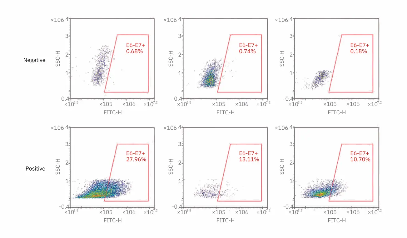
accurate detection of HPV E6/E7 mRNA (Figure 2). The threshold is set at 2% of cells within the E6/E7 positive analysis gate, which was determined by previous studies done at Enzo Life Sciences. All HPV positive samples had more than 2% of cells in the analysis gate, while all negative samples made up less than 2% of the E6/E7 gate.
Figure 2. E6/E7 mRNA expression detection in known negative and positive ectocervical cells. Cytology samples were evaluated for the presence of E6/E7 mRNA. Representative positive and negative samples are shown. Threshold is 2% cells within the E6/E7 gate. All HPV negative samples were <2% within the E6/E7 analysis gate, and all HPV positive samples were >2% of the E6/E7 gate.
Conclusion
Detection of HPV mRNA detection in cervical samples is a direct indication of viral activity. With the use of the FLOWSCRIPT HPV E6/E7 assay and the NovoCyte flow cytometer, analysis of HPV RNA transcripts can be assessed on a single-cell level even in mixed cell populations. Measuring transcripts for HPV RNA is easy on the NovoCyte flow cytometer. Hands-on time is minimized by the use of an optional autosampler (NovoSampler Pro), which can automatically analyze samples in tubes or plates. Due to the wide, dynamic range of detection, there is no need for PMT voltage adjustments limiting the variability between users. With the NovoCyte flow cytometer, HPV RNA detection results are fast and easy to obtain.
References
1. Saslow, D; et al. American Cancer Society, American Society for Colposcopy and Cervical Pathology, and American Society for Clinical Pathology Screening Guidelines for the Prevention and Early Detection of Cervical Cancer. J. Low Genit. Tract Dis. 2012, 16(3), 175–204.
2. Walboomers, J. M.; et al. Human
Papillomavirus is a Necessary
Cause of Invasive Cervical Cancer
Worldwide. J. Pathol. 1999, 189, 12–19.
4. de Sanjose. S.; et al. Human Papillomavirus Genotype Attribution in Invasive Cervical Cancer: a Retrospective Cross-Sectional Worldwide Study. Lancet Oncol. 2010, 11, 1048–1056.
Wright, T. C. Jr.; et al. Evaluation of HPV-16 and HPV-18 Genotyping for
the Triage of Women with High-Risk HPV Positive, Cytology-Negative
Results. Am. J. Clin. Pathol. 2011 Oct, 136(4), 578–586.
For Research Use Only. Not for use in diagnostic procedures.
This information is subject to change without notice.
© Agilent Technologies, Inc. 2019 Printed in the USA, November 1, 2019 5994-1025EN AN 15 DE.3974305556
Invite our experts and lift your research to the next stage with a demo of Novocyte from OLS
Your Partner in Cell Research
Request Cell Novocyte Demo